

Portable Ultrasound Device Supports Early Detection of Breast Cancer
SadaNews - Researchers at the Massachusetts Institute of Technology have developed a portable ultrasound imaging system aimed at making breast screening more accessible and regular, especially for women who are at a higher risk of developing breast cancer or those with dense breast tissue. While the system does not replace established medical screenings directly, it introduces a new concept for how to monitor breast tissue more frequently, either in the clinic or potentially at home in the future, without the need for a specialized operator each time.
Gap Between Annual Screenings
Many early detection programs rely on mammography once a year. However, some tumors may develop between screenings, known as interval cancers. According to a report from "MIT", these cases represent between 20 to 30 percent of breast cancers and are often more aggressive.
This concern led Kanan Dagdevren, an associate professor of Arts and Media Sciences at "MIT" and the principal investigator on the study, to develop an imaging method that could be used more frequently than traditional mammograms and is particularly suitable for women with dense breast tissue. Dagdevren lost a relative to cancer that developed between two annual screenings, prompting her to think of a technology that could support closer monitoring.
Imaging from a Small Device
The new system relies on a small ultrasound probe connected to a data acquisition and processing unit that is only slightly larger than a smartphone. This system can create a three-dimensional image of the entire breast by scanning only two or three points, rather than relying on large machines and specialized operators as is the case with many traditional ultrasound screenings.
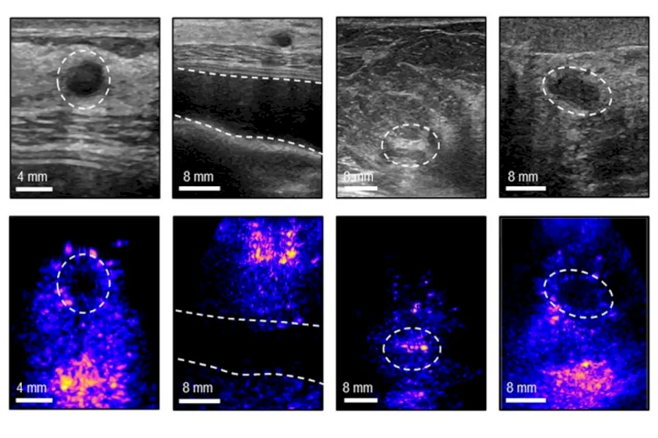
The new study, published in the journal "Nature Communications", indicates that the team improved the image quality and accuracy compared to previous versions. Researchers added a backing layer to the ultrasound transducer, helping to contain and focus the waves, and reduce acoustic and electrical noise, leading to clearer images. The team also developed an algorithm to adjust the formation of the sound beam according to the varying speed of sound in different tissue types, such as skin and fat. The researchers noted that this processing improved accuracy by up to 10 percent.
User-Guided Interface
The significance of the project goes beyond miniaturizing the device or improving the image; it extends to usability. The researchers designed a computer interface that guides the user in placing the probe in the correct location while displaying live images on the screen. Dagdevren states that the interface helps place the device in the same location for each exam, which is important for monitoring the same tissue over long periods.
The team tested the ability of non-specialists to use the system. In a trial with ten volunteers, they were asked to identify small targets within a model simulating human tissue, and they achieved better results using the new system compared to a traditional probe. In another trial involving seven individuals, participants managed to correctly position the probe during repeated examinations.
Monitoring Treatment, Not Just Diagnosis
This type of device may play a role in early detection, but it could also be important in tracking patients after treatment or during it. The ability to image the same site multiple times can assist in monitoring known changes, such as fibroadenomas or microcalcifications, or tracking a patient's response to pre-surgical treatments.
The researchers believe the technology may also reduce the full reliance on the availability of ultrasound technicians, especially in areas lacking specialized personnel. The team hopes to develop an interface that could potentially be used on a smartphone or tablet in the future, making the system more portable and suitable for daily use.
Research Step Before Commercial Application
Despite the promises of the system, it remains in the research phase and has not yet transitioned into a commercially available medical device. Some team members are working to establish a company to push the technology toward practical use, considering breast imaging as the first targeted application. Dagdevren points out that the platform could later extend to imaging other soft tissues, such as monitoring ovarian cancer, measuring the development of the uterine lining, or monitoring the fetus.
The project represents a growing trend in portable medical devices, transferring part of imaging capabilities from hospitals to smaller, more user-friendly tools, without sacrificing accuracy or medical follow-up. If this technology successfully passes testing and regulatory phases, it could bring breast imaging closer to women who need frequent monitoring rather than just annual screenings.
 Follow the latest Sada News via Google News
World Cup: Spain Advances to Semi-Finals with Late Victory over Belgium

Meta Prepares to Produce Custom AI Chip to Double Its Computing Power

Portable Ultrasound Device Supports Early Detection of Breast Cancer

15 Foods That May Keep Your Body Hydrated Besides Drinking Water

What Happens to the Kidneys When You Consume Energy Drinks Daily?

Ear Acupuncture for Relieving Migraine Pain

Sunscreen... What is the appropriate amount? And how to choose the best product?
